BA Festival of Science
1. Details of presentation:
Of the five senses our sense of touch is the only one that is not located only in your head (as are sight, hearing, taste, smell) – and near to your brain – but is found all over the body in the skin, making it our largest sense organ. Its sensory receptors are found in the two main layers of the skin – the epidermis and dermis – and in the muscles and joints, and is collectively known as the somatosensory system. In this presentation we will talk only about the sensors found in the skin – the cutaneous sensory system – those found in the muscles and joints are called proprioceptors and convey messages about deep pain and body/limb position to the brain. The skin has millions of nerve fibres that until relatively recently had been broadly categorised into three main types, based on the known cutaneous senses they serve of touch , temperature and pain/itch – and these are further subdivided into about twenty different subtypes of nerve endings and receptors that tell you if something is hot or cold, prickly or soft, stinging or burning etc.
However, in this presentation we will describe not only research we have done investigating the ‘classical’ cutaneous sense of touch, employing a range of new techniques, but review research we have carried out in the past 10 years that has discovered another cutaneous sensory nerve fibre system in human skin that appears to code for the pleasant and affiliative aspects of touch we are all familiar with, such as when grooming, or being cuddled.
Before describing this new system we need to know a little more about the nerve fibres that innervate the skin. There are three main types that are distinguished by how fast they conduct – like a wire – bioelectric activity to the brain. Two of the three types are ‘fast’ conducting nerves – called A-fibres – and are covered in a thin fatty sheath (called myelin), like the insulation around a wire, which helps them achieve their high conduction velocities. This is why when something touches you, you feel it immediately – essential for manipulating tools and handling objects. The third type, however, called C-fibres, have no myelin sheath and therefore send signals to the brain very slowly. It is these nerves that are probably one of the most important to us as they serve the sense of pain – importantly there is also an A-fibre pain system (called ‘first pain’) that responds very quickly to a painful stimulus, which is why when you touch a hot stove for example you pull your hand away reflexively before you really think about what has happened, but you know in a few seconds a deep throbbing and burning pain is going to strike – this is the brain’s response to C-fibre activity (called ‘second pain’) coming from the skin, and we all know how emotionally distressing and unpleasant this kind of pain can be. Different parts of the body are more or less sensitive than others because they have more nerve endings. If you get a piece of grit in your eye, have a toothache, or bite your tongue, it hurts so much because there are more C-fibres there. The research we and our academic colleagues in Sweden have been doing is building evidence for another role for C-fibres in the skin that are not pain receptors – nociceptors – but are pleasure receptors – hedonoceptors, and in a similar way to touch and pain sensitivity being different across the body, our research is showing that sensitivity to pleasant touch is also highly heterogeneous – some areas like being touched more than others!
Employing a sophisticated nerve recording technique (microneurography), where very fine tungsten microelectrodes (a bit like acupuncture needles) are inserted through the skin and into an underlying nerve bundle, we have been able to record the electrical activity from many of the different nerve fibres types described above, particularly the mechanosensitive ones. It was during such research that a new class of touch mechanosensitive nerves was discovered in (Nordin, 1990) that were not the usual fast conducting, classical touch A-fibres, but were slowly conducting C-fibre nerves, which had up until then only been thought to respond to painful stimuli. The type of stimulus that most excited them was a slowly moving gentle stroking one, such as that delivered during a caress or when being gently massaged! These light-touch sensitive C-fibres are known as C-tactile nerves, or CT’s and one of their defining features is that they are not found in glabrous skin (the palms and soles of the feet), but only in hairy skin (this term is used to describe all other skin sites on the body). We are now understanding touch in a similar way to how we understand pain, in that there are two types of touch, as there are two types of pain – ‘first touch’ subserved by fast conducting A-fibres, and ‘second touch’ subserved by this new class of mechanosensitive slowly conducting C-fibres.
In order to study ‘first’ and ‘second’ touch we have employed a range of cognitive neuroscientific techniques from areas of neuroanatomy, psychophysics, neuropharmacology, neuroimaging, cognitive science and affective neuroscience, and have developed precision stimulators that allow us to excite first and second touch nerves. Experimental results from two in particular, the Piezo Tactile Stimulator (PTS) and the Rotary Tactile Stimulator (RTS), will be described in this presentation.
The PTS allows us to deliver highly controlled vibrotactile stimuli to the skin of a subject, usually to the digit skin , during a form of neuroimaging called functional magnetic resonance imaging (fMRI), that measures local changes in blood flow in the brain that are correlated with activity in specific areas – we can literally see inside the sensing and thinking human brain. As fMRI uses very high strength magnets no metal objects can be taken into the scanner so we needed to develop a tactile stimulator that was non-metallic – the PTS – and that could deliver precisely controlled vibrations to the skin that ranged from microns in amplitude to millimetres – your sense of touch is exquisite, just blow gently on your hand and you will see just how sensitive the mechanoreceptors in your skin are. What is unique about our touch-fMRI studies, carried out at the Sir Peter Mansfield Magnetic Resonance Unit in Nottingham (where the Nobel prize for fMRI was recently awarded), is that we have not only been able to stimulate touch receptors, very precisely, but we are the only group in the world so far to have used the microneurography technique inside an fMRI scanner, where we have electrically micro-stimulated a single touch nerves and measured the subsequent activity in the brain’s somatosensory cortex. The electrical micro-stimulation of a single touch nerve causes a ‘phantom touch’ perception where you are convinced something is gently pressing into your finger – this is akin to a well known and very distressing form of pain called ‘phantom limb pain’, and research such as our may help to understand the causes of this often untreatable pain condition, as well as furthering our understanding of the sense of touch.
We developed the RTS to allow us to excite CT’s across the body surface and to study their responses using psychophysical measures [psychophysics is a psychological procedure that allows the study of the relationship between the physical aspects of a sensory stimulus and its subjective or perceived correlates], and electrophysiological measures [the direct nerve recording technique of microneurography]. We will show that people’s subjective responses to being gently stroked by our touch robot, and the responses of their CT nerves, follow the same response profile. Namely, CT nerves are most excited by stroking velocities around 3 – 5 cm/sec, and low forces of about 1gram, and it is just these stroking velocities and forces that people report in our psychophysical studies as being most pleasant!
The evolution of a system of cutaneous C-fiber nerves that respond to both pain and pleasure is seen as fundamental to survival. With pain it has been clearly established that without such a sense we would not survive, and now we are beginning to understand that without a sense of ‘pleasure’, or as we prefer to call it ‘reward’, behaviours that we take for granted like the caress between lovers and the nurturing of babies – all driven by skin-to-skin contact – we would also not survive……
2. Key finding of the research:
The use of the converging methodologies of microneurography, psychophysics and neuroimaging, plus a multidisciplinary team of scientists, has enabled us to discover and characterise the neurophysiological, psychological and emotional properties of a fourth dimension in the classical skin senses that now includes pleasure, as well as touch, temperature and pain/itch.
The demonstration that we can couple microneurography with fMRI has opened up a whole new field of human research that will not only obviate the need to use animal models in neuroscientific research, but as importantly allows us unrivalled access to the study in humans of somatosensation. There are many debilitating diseases that affect the peripheral nervous system such as diabetes, Aids, neuritis, neuropathy, carpal tunnel syndrome etc. and our research is providing new ways of understanding and potentially treating such conditions.
The tuning properties of CT’s shows that they respond optimally to specific stroking velocities of approximately 4 cm/sec and forces of around 1 Newton, and less so to faster and slower velocities, or higher or lower forces, and that the response profile of these nerves matches precisely the subjective perceptual reports, as measured psychophysically. Additionally, neuroimaging studies with fMRI and PET have corroborated both these findings, showing that the brain areas that respond to such pleasant forms of touch are those areas that are known to process emotions – of both pain and pleasure.
3. New and interesting aspects of this research:
The description of a ‘new’ touch modality in human skin, one that shares the same nerve type as pain, is seen as of potential relevance to not only a better understanding of the role of touch in human social behaviour and personal well-being but also to a better understanding of human pain mechanisms and their treatment. We have new evidence that stimuli that excite CT’s, reduces activity in pain C-fibres. We are also interested in studying a range of clinical conditions, from depression to autism, that are also known to have links with touch – most autistic children hate being cuddled and stroked, and many depressed people show clear signs of lack of body care, such as lack of grooming behaviours, and a susceptibility to depression may have its roots in poor maternal care and early life experiences with touch starvation.
And at a social level, the description of an affiliative and affective (rewarding) tactile system will enable us to better understand not only the importance of this sense in child development, but also its role in adolescent behaviour, where the pleasure gained from intense tactile interaction, such as during an E-fuelled rave, may be driven by the effect this drug has on serotinergic systems in the brain which we have shown can effect responses to touch. The importance of touch in ageing is also of great interest – many elderly people live is isolation and therefore do not get adequate access to affiliative or affectionate touch – one reason why owning a pet can be so palliative, or a visit to the hairdressers – we have shown with fMRI that stroking the scalp activates all the brain’s pleasure networks…….
4. Relevance to a general audience:
Most people, when thinking about their senses, will generally consider their senses of sight and hearing as the most important to them. What we will describe to them with our recent research into the sense of touch will help them consider the importance of this sense in those aspects of their lives that relate more to affective and nurturing behaviours – such as how they interact with their partner or children, or grandma or granddad. The gentle touch on the shoulder can convey more when helping a distressed friend than words can ever convey, and ‘touch starvation’, we are beginning to learn, could have dire social consequences later in life, as well a adverse effects on health and mental well-being.
5. The next steps:
The description and characterisation of affective touch mechanism in human skin is at a very early stage. So far we have identified a specific class of peripheral nerves subserving second touch, and have exposed some of the brain areas that that process this form of touch. We have little knowledge of the receptor mechanisms operating at the sensory encoding stage i.e. the neurobiological mechanisms that are responsible for transducing mechanical touch into a neural signal, for either the first or second touch systems. Research into skin sensory receptor mechanisms has advanced most in the field of pain research, notably with the discovery of a class of receptors called transient receptor potential (TRP) receptors that are found in peripheral nerves endings responsive to temperature and pain, and we plan to extend our research to study the neurochemical mediators of second touch at both the peripheral and central nervous system levels.
We are also aiming to investigate the social basis of touch, particularly its role in affiliative (nurturing) and grooming behaviours. We see human grooming behaviours as more than simply functional i.e. maintaining the physical health of the skin by removing micro-organisms and dirt, but as importantly it generates feelings of well-being, and it is this ‘reward’ value that drives the behaviour – we do more of what we like.
6. Relevant publications:
Nordin, M (1990) Low threshold mechanoreceptive and nociceptive units with unmyelinated C-fibres in the human supra-orbital nerve. J. Physiology 426: 229-240
Essick, G K, James, A & McGlone F P. (1999) Psychophysical assessment of the affective components of non-painful touch NeuroReport 10
Francis, S., Rolls, E., Bowtell, R., McGlone, F., O’Doherty, J. & Smith, E. (1999) The representation of the pleasantness of touch in the human brain, and its relation to taste and olfactory areas. NeuroReport 10, 453 – 459.
Olausson H, Lamarre’ Y, Backlund H, Morin C, Wallin BG, Starck G, Ekholm S, Strigo I, Worsley K, Vallbo AB, Bushnell MC. (2002). Unmyelinated tactile afferents signal touch and project to insular cortex. Nat Neurosci 5: 900-904
McGlone F, Kelly E, Trulsson M, Francis S, Westling G, Bowtell R. (2002) Functional Neuroimaging Studies of Human Somatosensory Cortex. Behavioural Brain Research 135(1,2): 147 – 158
McGlone F, Olausson H, Vallbo A & Wessburg J (2007) Touch and Emotional Touch Canadian Journal of Experimental Psychology( in press)Olausson H, Cole J, Rylander K, McGlone F, Lamarre’ Y, Wallin G, Krämer H, Wessberg J, Elam M, Bushnell C & and Vallbo A (2008) Functional role of unmyelinated tactile afferents in human hairy skin: sympathetic response and perceptual localization. Experimental Brain Research 184(1): 135-140
McCabe C, Rolls ET, Bilderbeck, A and McGlone F. (2008) Cognitive influences on the affective representation of touch and the sight of touch in the human brain. Social Cognitive & Affective Neuroscience (available online)
Harlow, H F. (1958) The nature of love. Am Psychol. 13: 673- 685.
Bessou,P, Burgess, P, Perl, E & Taylor, C (1971) Dynamic properties of mechanoreceptors with unmyelinated C-fibres. J. Neurophysiol. 34: 116-131
8. Media links:
read moreMultisensory Product Design
There has been a growing recognition that developing a new product is still a largely fractionated process, with different groups working on different aspects of the product from the formulation chemist to the package designer, from the consumer evaluator to the marketer. It is important to integrate this process, with respect to a product’s sensory properties, to ensure that these are engineered into all stages of development.
How can we engineer ‘liking’ into products in a predictive rather than trial and error way? How can we find out what it is that people like about using products? How can we better measure ‘liking’? How can we measure behaviour without disrupting it? How can we give people pleasant experiences that they cannot themselves articulate they want from our products? These and other questions can be raised and addressed in a workshop format that NeuroSci can organise.
FMCG companies make a wide range of products, from soap powders to soups, and although these products have particular functional benefits that may drive purchase decisions, their selection in a ‘shopper’ context is dependent on a number of factors that precede (from days to seconds) actual decision to purchase, and these drivers are often associated with a wide range of sensory cues that are not available to conscious interrogation. The sensory / perceptual / emotional properties of these everyday products, as well as the influences from advertising and other forms of communication, play a vital part in their selection and use, over a competitor’s product that will often perform equally well – functionally. However, to engineer products that will be selected predicatively is still a largely empirical and iterative process, reliant upon verbal self-report as the primary ‘measurement tool’. Recent developments in the social, cognitive, behavioural and neurosciences has led to a new understanding of the ways in which we perceive and interact with our environment, and many of these advances are of relevance to the design of the everyday products.
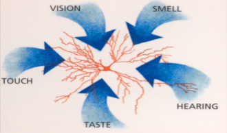
One particular field in Cognitive Neuroscience that is of particular relevance to product design is multisensory perception, where it is now known that the primary senses of touch, vision, smell taste and hearing are processed in the brain, not as single sensory modalities, but as mixtures, and are ‘fused’ together to form our perceptions and inform our actions. These processes are largely hidden from our conscious experiences and operate at an implicit and often automatic level. For example the smell of an object can be changed by its colour, its feel by its sound, or its selection by the shape of the package. Even though we might intuitively know that ‘X’ goes with ‘Y’ in a particular product format, we still do not KNOW how to predict the sensory or emotional performance of a product, and too often rely upon an iterative, costly and ‘noisy’ process of asking consumers if they like it. There are better ways to build better products………
read moreIce Cream makes you happy
Changes in brain activity related to eating Carte D’Or ice cream
1. Background and Rationale
Recent research has employed brain imaging technologies to do two things of interest to us: (1) to investigate the neural underpinnings of the reward value of different food stimuli (how our brains determine if something is pleasant or pleasurable or not)
(2) to try and understand why a consumer responds or behaves in the way they do without having to use verbal report (which only provides information of which the consumer is aware and may be rationalised to make it more understandable to that consumer or the person asking).
Other brain imaging research has shown that there is an area of the frontal brain (the orbitofrontal cortex or OFC) that seems to be involved in representing the emotional aspects of food products. The OFC is activated when there is a reward value to the stimulus i.e. a pleasurable or painful stimulus, these stimuli activating different areas within the OFC. The OFC does not respond to mundane stimuli (e.g. ordinary food). In fact it you damage the OFC people’s emotions are neutered.
We know from what consumers tell us that ice cream is considered a highly pleasurable food, is often eaten as a treat or indulgence, has unique and very pleasing sensory properties and is strongly associated with positive memories and experiences. This is what makes people want to eat ice cream. While we strive to improve the nutritional profiles of our products (for consumption as part of a balanced diet) it is essential that we maintain this pleasurable experience and also to show that the pleasure itself makes us happy, which in turn is beneficial for our health.
To measure objectively whether ice cream is pleasurable, and therefore makes us happy, we can use brain imaging to see whether ice cream activates the pleasure/reward centre of our brains (the OFC).
2. Goal of the Study
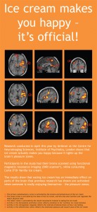
The goal of this study was to provide scientific evidence that “ice cream makes you happy” by demonstrating that the OFC (the pleasure/reward centre of the brain) is activated by consumption of Carte D’Or ice cream, using fMRI brain imaging.
3. Research Investigators
The study was run at the Centre for Neuroimaging Sciences, Institute of Psychiatry, London in collaboration with Professor of Neuroimaging, Michael Brammer. The study was designed and overseen by Unilever’s cognitive neuroscientist Professor Francis McGlone and managed by Dr Amanda Mistlin.
- Study Design
4.1. Participants
8 graduate and post-graduate staff at the Institute of Psychiatry participated in the study. They were chosen because they were all familiar with being in the scanner for the time needed (i.e. wouldn’t be anxious and would provide usable brain images), and were all happy to eat ice cream. None of them had any prior knowledge of this study.
4.2. Protocol
Prior to going into the scanner the participants were talked through the protocol and laid on the ground to receive a trial ice cream sample (to mimic how they would receive the sample in the brain imager). They were asked to rate how much they liked the ice cream on a 5-point scale (from +2 to -2).
In the scanner room the participant was laid down on the scanner table, with headphones on, and with their head encased in a frame. The frame acts as a radio receiver to sense the signals emitted from the brain. The participant is then slid into the bore of the fMRI scanner and given a preparatory scan.
Then the experiment with the ice cream begins. The participant is given a pipette of warm water, directed into their mouth (to cleanse/clear their palette). Thirty seconds later they receive their first sample of ice cream (~2g). The ice cream is delivered on the handle of a teaspoon by someone leaning into the bore of the scanner, and placing the end of the teaspoon between their lips. The aim is to do this with no head movement of the participant as this interferes with the scanning signal. The subject was advised to let it melt in the mouth as naturally as possible and then gently swallow. Each participant received 15 samples of ice cream in total, each 1 minute apart. Every 30 seconds after ice cream delivery the subject received a pipette of warm water to cleanse their mouth. This water acted as the control stimulus for the ice cream.
At the end of the ice cream feeding trial the participant is again scanned to get an anatomical view of their brain onto which the brain activation is mapped.
The whole session took approximately 30 minutes. At the end of the session the participant was again asked to rate how much they now liked the ice cream.
4.3. Products
Carte D’Or Vanilla Ice Cream, stored and served at the recommended temperature, was used in this study.
4.4. fMRI Scanner
We used a 3T (Tesla) scanner for this study to ensure sufficient power and resolution to see the areas of interest in the brain.
fMRI works by detecting local changes in blood flow in areas of the brain that are engaged in some form of mental event. The brain’s cells – neurones – are very dependent upon a good supply of nutrients, so when a specific area is processing a particular sensory input, e.g. the taste of ice cream, cells in the primary taste cortex increase their activity, thereby causing a local increase in blood supply to that specific area of the brain.
Subjects participating in fMRI experiments are placed inside very strong magnets, which literally magnetise them, polarising all the hydrogen nuclei (we are mostly water) so that their protons are aligned with the magnetic field of the magnet. Using radio frequency pulses (RF) these protons are briefly kicked out of alignment and when they relax back to their pre-disturbed state, emit a tiny RF signal. This is detected, localised by some very clever software analysis and eventually reconstructed to give a picture of the brain that reveals where the activity related to the stimulus was occurring.
When the brain is scanned a full volume image of the brain is created. This is sliced like salami into approximately 12-15 slices. This is done 3 times to get 3 volumes with 36 or more slices. This slice is further divided up into squares (voxels). Each one of these registers any change in the MR signal which is produced by the RF pulses and depends upon the level of deoxygenation in the vasculature within that voxel. If the neurones are active, the blood flow goes up, the balance of oxy to deoxy changes and you get, or don’t get, a signal change – the haemodynamic response function (HRF). You look at the HRF’s in particular brain areas correlated with the stimulus-on period (when ice cream is present) and subtract the no-stimulus condition (water) – using statistical t-tests you see which areas were significantly activated when your stimulus was present. The analysis was carried out using a statistical package written by Professor Brammer.
- 5. Results
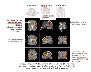
What we were hoping to find, and did find, was that eating ice cream activates the part of the brain called the Orbitofrontal cortex (OFC), which indicates the positive emotional pleasure and reward value of the ice cream.
We also found activation in other areas of the brain:
- The primary somatosensory cortex – activated by the texture and temperature of the ice cream
- The insula cortex – activated by the taste of the ice cream. This area also monitors and regulates the body’s physical state.
- The motor cortex – activated by the mouth movements involved in eating this ice cream.
- The dorso-lateral prefrontal cortex. Activity in this area reflects attention to the ‘exciting’ ice cream stimulus.
- The retro-splenial cyngulate. Activity here reflects the internal emotional check on how I am feeling.
The following brain image slices show this activation.

- Conclusions
Carte D’Or ice cream does light up the pleasure centre of the brain. It also activates other areas of the brain involved in sensory processing of the stimulus as expected, and activates areas of the brain involved in attention and processing of emotionally relevant stimuli.
Does pleasure equal happiness? Yes, because according to the field of Positive Psychology, happiness is simply a measure of positive feeling or emotion.
Do other foods stimulate the same area? Other foods with equally sensational sensory characteristics and a strong emotional value might light up the OFC, but we haven’t measured them. In fact, it is pioneering research to actually test foods with an fMRI scanner because typically you have to avoid any head movements associated with chewing. Ingestive behaviour is technically very demanding. We have been able to look at ice cream only because it is a semi-solid food, and because of the clever analysis available to compensate for stimulus-correlated motion. There has been a PET research study (Positron Emission Topography), not fMRI, using chocolate where activation of the OFC was found. Chocolate too is a unique combination of sensory characteristics, with melting properties like ice cream, with strong emotional valence. We have the benefit of combining ice cream and chocolate in some of our products – two pleasure zone activators – which maybe explains why this combination is so loved.
read moreNegative Skin Sensations
Many marketed body-care products are known to cause irritation, usually only in a small sub-population, but can result in a product’s withdrawal. Identifying these populations and the underlying mechanisms that case negative skin sensations is critical. Irritation manifests itself in a number of different ways, but broadly speaking these are either purely visual or purely sensory, or more often both. Visual irritation manifests as changes in the redness, dryness and swelling of the skin, without any sensory consequences, whereas sensory irritation is accompanied by the perception of sting, itch, burn, pain and tightness. Visual irritation accounts for approximately 33% of irritation, with sensory irritation accounting for the remaining 67%. Currently, 70% of women and 50% of men perceive that they have sensitive skin, but this self-perception is known to be highly unreliable.
Irritation is caused by molecules such as alpha hydroxyl acids (AHAs), retinol, perfumes and surfactants. The type of irritation that is caused by these molecules is not uniform and different molecules cause different types of irritation in different populations. This highlights the needs for a multifactorial approach to the study of irritation and a range of capabilities to measure & predict its different aspects.
Negative skin sensations are known to be affected by the irritant molecule itself, overall formulation, global region, ethnic group, body site and genetic factors. Complaints of irritation can also prevent commercialization of new molecules, resulting in the levels of actives that are known to be effective having to be reduced in formulations – even though consumer complaints may not reflect a true population response. With ~70% of women and 50% of men perceiving that they have ‘sensitive skin’, and around 65% of women believing that this is caused by a reaction to a product, it is imperative that companies making such products know about the neuroscience of negative skin sensations. Our knowledge of skin neurobiology and our understanding of the receptors that mediate irritation, pain and pleasure have increased dramatically in recent years. NeuroSci knows about skin irritation and has excellent collaborations with researchers in the field – both scientists and clinicians – and has published a number of high quality research papers in this area.
read morePositive Skin Sensations
As with negative skin sensations, where a system of specific receptors and nerves in the skin tell the brain when the skin is being damaged, it is now also known that similar mechanisms of receptors and nerves in the skin also exist and tell brain about pleasant skin stimulation. A recent Nature Neuroscience paper on our work on positive skin sensations was widely reported in the media (links appended) and has generated a great deal of interest. Methods developed to study positive touch can be applied in a range of different areas of relevance to FMCG companies, from the feel of a new skin cream to the textural properties of a products packaging.
http://www.nature.com/neuro/index.html
http://www.google.com/hostednews/ukpress/article/ALeqM5gd0M0w77MTLXj8wzEOqtL-HQVolg
http://news.bbc.co.uk/1/hi/health/7992299.stm
http://www.dailymail.co.uk/health/article-1169575/Rub-better-fast-slow.html
http://www.theherald.co.uk/news/health/display.var.2501574.0.Pleasure_nerves_found_in_humans.php
http://www.express.co.uk/posts/view/94837/Proof-that-keeping-love-alive-s-no-stroke-of-luck
http://www.heralddeparis.com/scientists-find-pleasure-nerves/30540
http://news.scotsman.com/scitech/Secret-to-mindblowing-massage-Rub.5164608.jp
http://www.channel4.com/news/articles/society/health/humans+hard+wired+for+sensuality/3084472
http://in.news.yahoo.com/139/20080911/936/thl-a-soothing-touch-really-does-make-pa.html
read moreThe Fat Duck
There have been many fruitful discussions over the past few years with Heston Blumenthal, who has a fascination with all things sensory – and as importantly he is keen to know more about how the brain deals with the multiple arrays of inputs coming in from the senses when we are eating; a truly multisensory behaviour. The pleasures we experience when dining – which are different in many ways from those we have when simply just eating – are all of course coming from the brain. Discovering more about how our brains’ process sensory signals from taste buds, olfactory, vision, hearing and touch receptors, and how our expectations and memories affect what we perceive when enjoying a good meal can lead to finding new ways to excite the senses and to enhance pleasure.
At the 2010 Cheltenham Festival of Science Heston and Francis talk about the pleasure brain and how he creates such wonderful dining experiences – http://cheltenhamfestivals.com/science-2010/hestons-sweet-shop/
read more


